Chapter 2 : The Spinal Cord

Brain: Contents Page

Sensory Neurones (' Afferents')

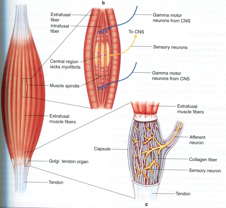
Sensory nerve endings can be classified according to morphology (Encapsulated vs Free nerve endings) or by their functional properties.

Sensory receptors transduce tactile stimuli and signal the intensity and temporal changes in the forces applied to the skin by passing trains of action potentials to the spinal cord. Each single afferent axon provides information about events in the area of skin it innervates (the receptive field) and the position and direction of movement of a tactile stimulus are computed from the patterns of activity in many axons, as the stimulus moves from the receptive field of one neurone to another. There are several different types of nerve ending in the skin which each respond to different, specific aspects of the stimulus and there is a good correlation between the function and structure of these nerve endings.

In skeletal muscle, there are also specific receptors that monitor the changes in length and tension of individual muscles, and there is also a good correlation between the function and structure of these nerve endings.


Structure of Sensory Nerve Endings

Location of Nerve Endings
Encapsulated Nerve Endings
Free Nerve Endings


Functional Properties of Sensory Receptors.   Top

Adequate Stimulus

Electrical recordings of action potential trains in single afferent nerve fibres ('single units') show that the information carried by each sensory axon relates to a specific type of natural stimulus ('modality' - mechanical, thermal, etc); their transducers are fairly specific for a particular form of natural stimulation. The 'adequate stimulus' is the specific natural stimulus (mechanical, thermal or noxious, etc) to which the transducers are most sensitive.

The adequate stimulus can be used to classify sensory receptors. Most sensory endiings respond to one main type of stimulus, such as light touch, stretch, or temperature. Thus for the sensation of touch, there are around four different types of sensory neurones responding to:

  • indentation of the skin
  • lateral stretch of the skin
  • vibration (optimally at 30 Hz)
  • vibration (optimally at 250 Hz)

Others respond to hair movement, and the hair receptors are most highly developed in the vibrissae of animals such as cats.

Cutaneous receptors are classified into three main groups:

Mechanoreceptors respond to mechanical stress or strain, or vibration.

Thermoreceptors respond to the normal range of temperatures in the skin.

Nociceptors respond to damaging stimuli such as painful mechanical chemical or thermal stimuli. The skin proteins begin to denature around 45 degees centigrade, and heat nociceptors respond to temperatures above 45 degrees C.

The threshold stimulus is the intensity of the natural stimulus that causes the afferent endings to start producing action potentials. So the temperature threshold of thermoreceotprs is less than that of nociceptors. Also the threshold of touch afferents is less than that of the mechanical nociceptors that only respond to damaging stimuli.

The receptive field is the area of skin that is innervated by a single sensory neurone. Some receptive fields are small, and a few may be described as punctate. But many axons innervate a moderately large area of skin, and the receptive fields of single axons overlap with each other; the exact location of a stimulus is worked out by the CNS as a result of comparisons of the spatial distribution of activity in a large number of axons.

Overlapping receptive fields in the skin can also be demonstrated when a nerve is cut. The area of skin innervated by a single nerve trunk does not become insensitive following section of one dorsal root, because adjacent roots also innervate that area. Spatial discrimination may change, but the fact remains that the areas of skin innervated by single axons or nerve trunks overlap with others.

Adaptation

Rate of adaptation

  • A tonic (or slowly adapting) receptor is a sensory receptor that continues to produce action potentials throughout the duration of the natural stimulus. As a result the signals arriving at the CNS provide information about the duration and intensity of the stimulus. Some tonic receptors have a resting discharge and indicate a background level of the natural stimulus (such as temperature or stretch.
  • A phasic (or rapidly adapting) receptor is a sensory receptor that responds to the application or removal of a natural stimulus but action potentials do not continue throughout the duration of the stimulus. Instead, the activity reflects the rate of application of the stimulus, and the timing of movements. Phasic receptors are particularly adapted to monitor vibration, and the Pacinian Corpuscle is an example that responds to vibrations particulalry around 200-300 Hz. Other phasic receptor in skin respond optimally to frequencies of around 30 Hz.

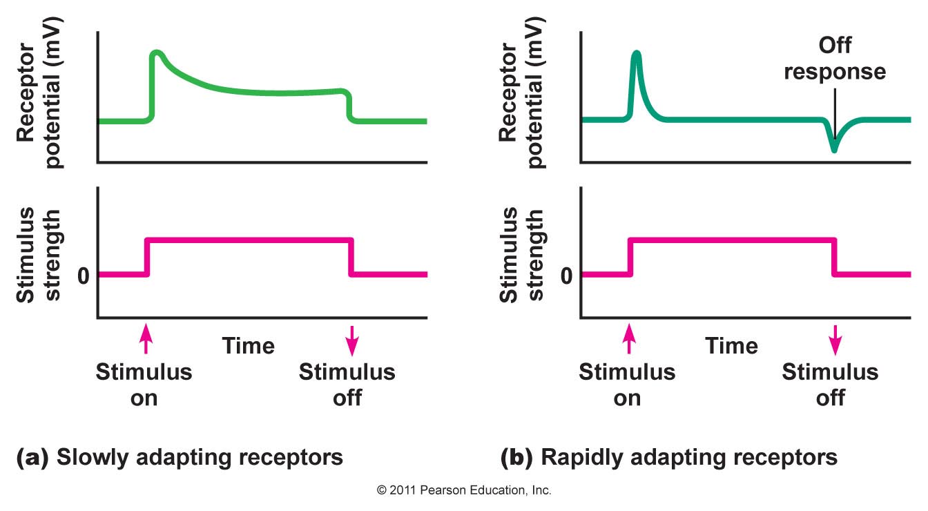
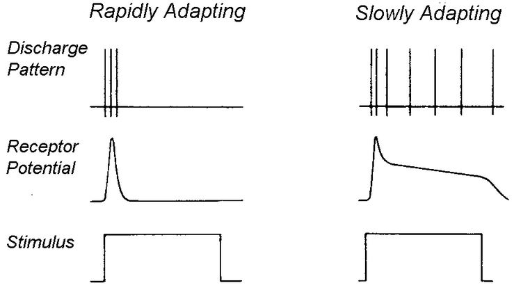
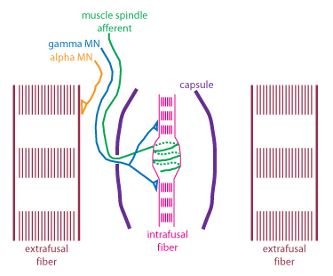
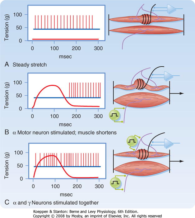
The receptor (generator) potential is the depolarising potential produced by the transducers at sensory nerve endings that initiates trains of action potentials in the afferent axon. It can be seen that in phasic (rapidly adapting) receptors the generator potential lasts only for the period during which the stimulus is applied.

It can be seen from the diagram that many slowly adapting receptors also show some phasic activity at the time the stimulus is applied. Whereas in slowly adapting (tonic) receptors the generator potential is maintained throughout the natural stimulus.

Some receptors monitor events inside the body : interoceptors include baroreceptors, chemoreceptors and osmoreceptors that sense arterial pressure, arterial blood gases or the osmolality of blood. This information is used to regulate the functions of internal organs, but does not enter consciousness.

Others, in muscle tendons and joints, are proprioceptors, responsible for the sense of kinaesthesia- the sense of position of the limbs.

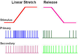
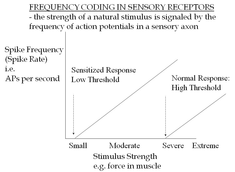
Frequency Code

The intensity of a natural stimulus is signalled by the frequency of action potentials generated during the tonic phase of their discharge.

In rapidly adapting receptors, the rate of discharge of action potentials is related to the change in the generator potential, which is dependent on the rate of application of the stimulus.

In slowly adapting receptors, the generator potential is maintained and that causes a train of impulses to occur in which the rate of discharge is related to the depolarisation. Most tonic, slowly adapting receptors, show some phasic activity related to the speed of onset of the stimulus.

*

Classification of axons according to diameter and conduction velocity   Top

Axons may be classified according to their diameter or their conduction velocity: for myelinated axons, there is a relationship between the two, and as a rule of thumb, the conduction velocity in m/sec is roughly the six times the axonal diameter.

The two forms of classification use different symbols: Groups A, B and C based on diameter, and Groups I, II, III and IV, based on conduction velocity. The former has been used in studies of skin nerves, and the latter in studies of muscle nerve afferents.

Classification by diameter : the A, B, C system

A fibres have large diameters, and C fibres are amall unmyelinated axons.
The A fibres are divided into four groups:

  • A alpha
  • A beta
  • A gamma
  • A delta

A fibres have diameters between 2 (A delta) and 20 A alpha
C fibres are unmyelinated (diameters of 0.2-1 micrometers)

Examples are ;

  • alpha motoneurones
  • gamma efferents
  • A-delta and C-fibre nociceptors

Classification by Conduction Velocity: the I, II, II, IV system

Myelinated fibres in Muscle afferent nerves are divided into four goups depending on conduction velocity

  • Group I - these are subdivided into Ia and Ib
  • Group II
  • Group III
  • Group IV

Group I have higher conduction velocities than the other groups, with Group IV being the slowest.

Examples are:

  • Ia - Muscle spindle primary endings
  • Ib - Golgi Tendon Organs
  • II- muscle spindle secondary endings
  • III- pressure pain receptors in muscle
  • IV- nociceptors
acbrown.com
Top

Muscle and Tendon Afferents

The Muscle Spindle

The muscle spindle is a small thin spindle-shaped encapsulated sensory organ which exists in most skeletal muscles in quite large numbers. The length of the spindle is just a few mm, and each end (or 'pole') is attached to the endomysium- the connective tissue surrounding the large skeletal muscle fibres that produce the powerful contractions. These powerful muscle fibres are known as extrafusal fibres, meaning that they are outside the spindle. In contrast, the muscle spindle contains some small short thin muscle fibres that lose their striations in the middle of the spindle, and these modified muscle fibres are known as intrafusal fibres (they are inside the spindle). Intrafusal fibres can contract and influence the tension and the dimensions of the central core of the spindle - the site where sensory endings wrap themselves around the central core.

There are two types of sensory ending in the muscle spindle - primary and secondary, with Group Ia and II myelinated axons respectively. These have similar functions - both are slowly adapting receptors that monitor the length of the muscle continuously - but with the primary ending having greater phasic activity than the secondary ending. In the diagram opposite only the primary ending is shown:- it spirals around the central core of the spindle, and anatomists call it the annulospiral ending. The secondary endings have different morphology and are located to each side of the annulospiral ending; they are known as flower-spray endings, because of a fanciful resemblance to a bunch of flowers!

 *


The phasic activity of muscle spindles has been used by physical therapists who use vibration to activate the endings and initiate reflex activity in the extrafusal fibres using the stretch reflex pathway.

Response of Muscle Spindle afferents to stretch

When a muscle is stretched, the muscle spindles are also stretched because it is attached to the endomysium and exists in parallel to the extrafusal muscle fibres.

So the response of the primary and secondary endings is to increase their rate of firing action potentials: the muscle spindle acts as a length detector, and the primary endings also show phasic activity..

Activity of Muscle Spindle afferents during Extrafusal Contractions

When extrafusal skeletal muscle fibres shorten, then muscle spindle is also shortened, and there is less activity in the afferent axons.

Again the muscle spindle acts as a length detector- this time detecting a shortening of the muscle.

Gamma Efferents

The intrafusal fibres at each pole of the muscle spindle receive a motor innervation from small diameter axons called gamma efferents, because of their size. Activation of the gamma efferents causes the poles of the spindle to contract, stretching the central core and distorting the afferent nerve endings. So the gamma efferents are capable of 'taking up the slack' within the spindle, and thereby adjusting its sensitivity to stretch.

The gamma efferents therefore are a mechanism by which the central nervous system can adjust the sensitivity of spindles in different conditions- a mechanism known as gamma bias.

It is known that during voluntary movements there can be co-activation of gamma motoneurones and alpha motoneurones, so the sensitivity of the spindle can be increased at the same time as the muscle shortens.

Top
More details on Muscle Spindles

Nuclear bag and nuclear chain intrafusal fibres
Gamma-static and Gamma-dynamic efferents


Golgi Tendon Organs

Golgi Tendon Organs are present in the tendons at each end of the muscle. They are relatively insensitive to forces applied to the tendon, and only become active when large tensions are generated by the muscle. These receptors are 'in-series' with the extrafusal fibres.

The nerve endings are entwined between collagen fibres within the tendon, and the receptor senses the average tension generated in muscle, just as the muscle spindle monitors muscle length changes.

Golgi Tendon Organs have Group Ib axons - slightly slower conduction velocities than the Muscle Spindle primary endings.

Classically the activity of Tendon organs contributed to the clasp knife reflex that can be elicited in some patients with neurological disorders. In this reflex, the tension applied to an active muscle is gradually increased, until the limb suddenly collapses, because tension receptors cause inhibition of those motoneurones.

Group III Muscle Afferents

Group III Muscle Afferents respond to strong applied pressure or stimuli approaching the injurious range to the muscle belly. They also respond to ischaemia and the accumulation of lactic acid in the tissues.

Their other name is 'pressure-pain receptors'.

Group IV Afferents

Group IV afferent fibres from muscle are concerned with sensing injury to the muscle. Damaged muscle can release various mediators that are involved in pain and are similar to those listed below as endogenous algesic agents that activate nociceptors in other tissues.

In muscles, tendons and joints, unmyelinated and finely myelinated afferents occur in the muscle, is connective tissue and and the synovial membrane. They generate action potentials in response to extreme stimuli - powerful forces, and extreme movements of joints

courses.washington.edu

Top


Skin Afferents

The mechanoreceptors (touch receptors) in skin can be classified as two types of slowly adapting and several types of rapidly adapting receptors, depending on the area of skin: glabrous and hairy skin have slightly different sets of rapidly adapting receptors.

The slowly adapting receptors are :

  • Type I - sensitive to indentation of the skin and mediated by numerous Merkel cells wrapped around the myelinated nerve endings
  • Type II - a receptor with some resting discharge that is modified by stretching the skin, possibly as a result of joint movement, and with a considerable sensitivity to changes in temperature. This type of receptor is associated with the Ruffini end organ.

Rapidly receptors include Pacinian Corpuscles, hair receptors, and (in glabrous skin) field receptors. There are several types of hair receptors, some of which are highly sensitive; in glabrous skin the field receptors, mediated by Meissner's corpuscles are also sensitive to light touch. Pacinian Corpuscles lie deep in the dermis and sense high frequencies of vibration.

Thermoreceptors are usually unmyelinated afferents. Type II slowly adapting mechanoreceptors are very sensitive to stretch of the skin, but are also sensitive to changes in temperature of the skin, so may also be involved in signalling skin temperature.

Thermoreceptors are particularly evident in the scrotum, and temperature changes to this area induce the cremasteric reflex, which controls the position of the testis, so as to keep it at an appropriate temperature to preserve fertility.

Nociceptors   Top

Unmyelinated axons occur in almost all tissues, and the sensory ones generally mediate the senses of pain and temperature. The sensory receptors that respond to injurious stimuli are called nociceptors. They induce reflex activity in flexor muscles and in the autonomic nervous system.

Nociceptors respond to injurious stimuli caused by

  • Heat
  • Pinprick and pinch
  • Chemicals
  • Cold

The mechanism by which nociceptors transduce noxious events involvesa superfamily of membrane proteins called TRPV receptors .

Transient Receptor Potential Vanilloid channels are a super- family of transient receptor potential (TRP) ion channels, that are selective for calcium and magnesium over sodium ions. Several members of the family are sensitive to elevated temperature, while others are sensitive to cold.

The TRPV super-family of transducers show sensitivity to not only to heat and cold, but some respond to :

  • acid conditions,
  • osmolality, and
  • capsaicin, a vanilloid molecule derived from hot red peppers.

Finely myelinated and unmyelinated afferents usually have no specific histological structures at their nerve endings. They normally generate action potentials only in response to extreme stimuli - powerful forces, and extreme movements of joints.  The Frequency Code indicates that the frequency of the action potentials signals the strength of the natural stimulus to the CNS; but the threshold of these receptors is high, responding only to intense stimuli.

Sensitization of unmyelinated afferent fibres

Sensitization occurs when an unmyelinated nerve sensory ending that is normally responsive only to extreme forces and movements, becomes sensitive to normal forces and movements.

One example of sensitization is inflammation: normally nociceptive nerve endings respond only to extreme forces and movements, but, once sensitized, they become much more sensitive and respond to normal movements. In both circumstances the messages they carry give rise to pain.

Endogenous Algesic Agents   Top

Inflammation

In an inflamed joint, a small movement can give rise to pain. In this condition, the unmyelinated nerve endings can become sensitized, and instead of responding only to large forces and movements, become very sensitive to any movement- because these nerve endings are affected by chemicals produced within damaged tissues, such as

    Bradykinin, and
    Prostaglandins

Chemicals with the ability to sensitize the nociceptive nerve endings are known as Endogenous Algesic Agents.

Some of the common over-the-counter analgesic drugs act by interfering with the production or action of these agents.

Neuropeptides are present in many unmyelinated nerves. Some neuropeptides can be released from sensory neurones and cause 'neurogenic inflammation', in which the afferent endings can become sensitized.

Bradykinin is a peptide produced by enzymes released from damaged cells acting on a plasma protein, and is found in fluid in inflamed joints. The effects of Bradykinin include :

    vasodilatation
    local oedema (extravasation of plasma proteins)
    pain
Prostaglandins (PGs) are released from injured cell membranes. Arachadonic acid is broken down to prostaglandins by an enzyme (cyclo-oxygenase) to produce
PGE, PGF2alpha and PGI. They have the following effects:

  • vasodilatation
  • increased capillary permeability to plasma proteins, - causing local oedema
  • sensitization of nerve endings - they are Algesic Agents

Fine unmyelinated afferents contain neuropeptides which can also sensitize sensory endings when they are released. These peptides attract cells of the immune system, cause vasodilatation and increased capillary permeability. These neuropeptides include:

    Substance P
    Calcitonin Gene-Related Peptide (CGRP)
Substance P appears to be a neurotransmitter in the pain pathway in the dorsal horn of the spinal cord.

Analgesic drugs

Some analgesic drugs act at peripheral nerve endings. Common analgesic drugs that are bought over the counter act by interfering with the production of prostaglandins that act on the peripheral terminals of unmyelinated axons.

These drugs include:

  • hydrocortisone and other more powerful corticosteroids (glucocorticoids)
  • Non-Steroidal Anti-Inflammatory Drugs (NSAIDs) including Indomethacin and Ibuprofen
  • Aspirin and Paracetamol

Peripheral Neuropathies

Neuropathies are disorders of peripheral nerves that give rise to symptoms such as pins and needles (paraesthesiae), pain and numbness in the periphery of the limbs.

Neuropathy can occur in metabolic disorders such as diabetes, or in nerve compression (say carpal tunnel syndrome), cancer, and as a side effect of drugs. There may also be signs of denervation of muscle (muscle wasting and loss of power)

Nerve Conduction is SLOWED in neuropathy: because of smaller axons, and loss of myelin
Summary

Nerve conduction velocity depends on axonal diameter, and the degree of myelination. In Neuropathy, nerve conduction is slowed due to axons becoming smaller in diameter and because of demyelination.
Large diameter sensory axons mediate touch, vibration and proprioception (position sense).
Pain is mediated by small diameter axons, particularly by unmyelinated axons. These sensory nerve endings normally respond only to intense stimuli, but can become more sensitive as a result of chemicals released near the endings.
Algesic agents sensitise small fibre afferents; their action is influenced by analgesic drugs such as aspirin, indomethacin and corticosteroids

Chapter 2 : The Spinal Cord

Brain: Contents Page

Top

© HumanPhysiology.Academy 2014-2015